This is
first of a series or articles which will examine nibs and
their shapes in detail. Bruce gives an introduction to the
Scanning Electron Microscope and shows some preliminary images
to give you an idea of what this equipement is capable of.
A Q&A will be run in conjunction with this article, to
participate please click on the link in the related links
box on the left..
Scanning
electron microscopes have been available since the mid 1960s.They
can achieve much higher magnification and have a better depth
of field than optical microscopes. This is an big advantage
for examining large curved things like nibs.
SEMs work
by firing a finely focused beam of electrons, in a vacuum,
at the sample. The beam is rastered sequentially across the
surface of part of the sample. Some of the electrons are reflected
back from the specimen, detected and displayed to form the
enlarged image direct on a computer screen, and captured digitally.
Sadly, electron microscopes only work with monochromatic electrons
(of one energy), so the pictures are inherently black and
white. There are techniques for later colouring the images.
As well,
the beam of electrons can be used to analyse non-destructively
individual points on the sample, for instance, to identify
the elements present in the "iridium" on the point,
and the differences in composition where the "iridium"
is welded to the gold of the nib. This is because the impinging
electrons interact with the elements present where the beam
hits. The gold atoms emit characteristic gold X-rays and the
silver and copper atoms emit silver and copper characteristic
X-rays, and these are proportional to the amounts of each
element present. The X-rays are collected by an X-ray detector
attached to the SEM.
A relatively
hard part of imaging the nibs is getting an evenly illuminated
image, without some sections being too bright and some parts
being in the darker shadows.
Chosing
the correct angle to show the aspects desired is also a challenge.
Logically,
an SEM in Australia can now be operated directly over the
Internet, or the images viewed remotely in real-time from
the US or anywhere. However, SEMs are common in Universities
and research laboratories, so there should be some in most
major cities.
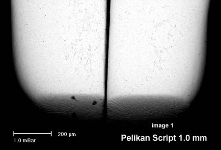
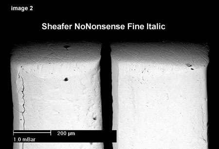
The illustrations
show some preliminary images from a recent Pelikan Script
1.0 mm low-price pen (sort of calligraphy) in image1, and
a standard Sheafer NoNonsense Fine Italic (which I bought
second-hand on Sunday for Aus$2. The Sheaffer is much squarer
("Italic") image2, and the Pelikan much rounder
and "Stub". The nibs are mounted upside-down, and
we are looking straight down at the underneath of the nibs.
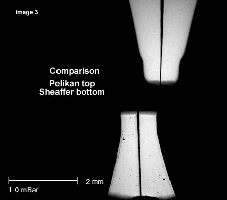
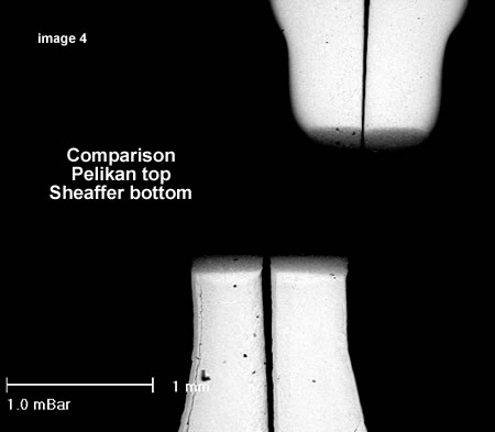
One advantage
of the SEM is that one can measure the dimensions relatively
accurately from the scale on the images(at least laterally
in the image if not vertically), and there is a good idea
of the shape, and the smoothness.I suspect that the very fine
scratches and features are not as important in nib-smoothness
as the actual shape and other factors. Obviously experts like
John Mottishaw who work with a good optical microscope will
have a very good feel for the parameters that matter.
|